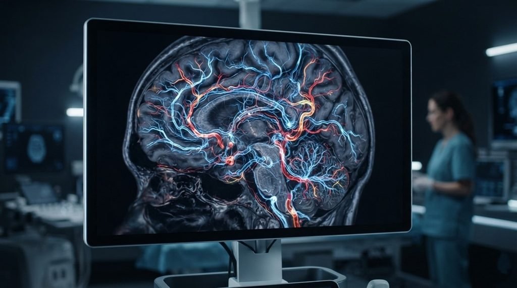
What if doctors could watch a stroke block blood vessels inside the brain in real time with perfect clarity?
🧠 Medical experts often struggle to see the microscopic blood vessels in the brain during the critical moments of a stroke event. Standard scanning tools lack the speed and sharpness needed to capture tiny, rapid changes in cerebral microvasculature. This visibility gap makes it incredibly difficult to understand exactly how tissue damage spreads when blood flow drops. Consequently, developing highly effective, targeted treatments for stroke victims remains a massive challenge for global healthcare systems.
🔬 A brilliant research team including Yoshihisa Kaneko and Takuro Ishii recently built a 4D cerebral microvascular imaging platform to solve this medical blind spot. This advanced system uses high-frame-rate ultrasound technology to map blood flow dynamics inside living brain tissues at a microscopic level. By capturing three-dimensional space over continuous time, the platform delivers real-time visual updates of deep vascular networks. The researchers successfully tracked immediate vessel changes in live models, proving that high-speed imaging can capture stroke progression as it happens.
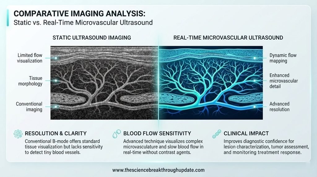
The Massive Blind Spot in Traditional Brain Scans
- ❌ Traditional imaging tools fail to capture the rapid, microscopic changes that happen inside cerebral blood vessels during a stroke.
- 📉 Standard scans lack the necessary combination of high spatial resolution and real-time speed to monitor deep brain networks.
- 🔍 Medical researchers cannot easily see how individual microscopic vessels collapse or adapt when a sudden blockage occurs.
- ⚠️ This lack of detailed data slows down the development of new, life-saving stroke therapies and emergency interventions.
How 4D Ultrasound Technology Unlocks the Brain
- 🛠️ The new platform developed by Tohoku University researchers utilizes high-frame-rate ultrasound to achieve unprecedented imaging depth and speed.
- 📊 This system records continuous three-dimensional data to create a true 4D map of active blood flow dynamics.
- 🔬 Scientists can now observe exactly how blood moves through vessels smaller than a hair during a simulated stroke.
- 🛡️ The technology provides a safe, non-invasive way to study brain circulation without relying on harmful radiation or contrast agents.
Why This Breakthrough Changes the Future of Medicine
- 🧪 Providing a clear window into the brain allows scientists to test experimental stroke drugs with incredible precision.
- 🏥 Doctors can better analyze how brain tissues recover, which opens the door for personalized medical treatments.
- 📈 The platform accelerates neuroscience research by showing how vascular networks react to various injuries and diseases.
- 🌍 This technology could eventually lead to faster diagnostic tools that save millions of stroke patients worldwide.
My Take: Watching the live collapse of tiny brain vessels changes everything. This practical tool cuts through medical guesswork, giving scientists the exact visual roadmaps needed to stop stroke damage instantly.
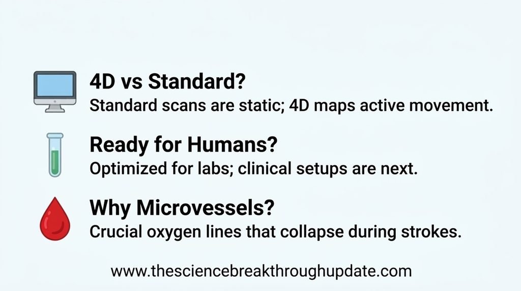
Reader Doubts: Core Questions Answered
1. How does 4D imaging differ from standard brain scans?
🖥️ Standard scans provide static, three-dimensional pictures of the brain. This new 4D ultrasound platform adds the element of real-time movement, allowing researchers to watch blood flow through tiny vessels continuously.
2. Can this technology be used on human patients right away?
🧪 This specific platform is currently optimized for laboratory research and live animal models. However, the engineering principles behind it are being developed to improve human clinical imaging systems in the future.
3. Why is microvascular health so important during a stroke?
🩸 Microscopic blood vessels deliver essential oxygen directly to brain cells. When a stroke blocks these tiny pathways, cells die within minutes, making real-time monitoring crucial for finding ways to stop the damage.
News source link: arXiv Quantitative Biology
Share this useful knowledge with your friends now!
Disclaimer: This content is for educational and informational purposes only.