Your bones are quietly conducting electricity right now, and a new scientific breakthrough explains how tracking these currents could stop osteoporosis before it starts.
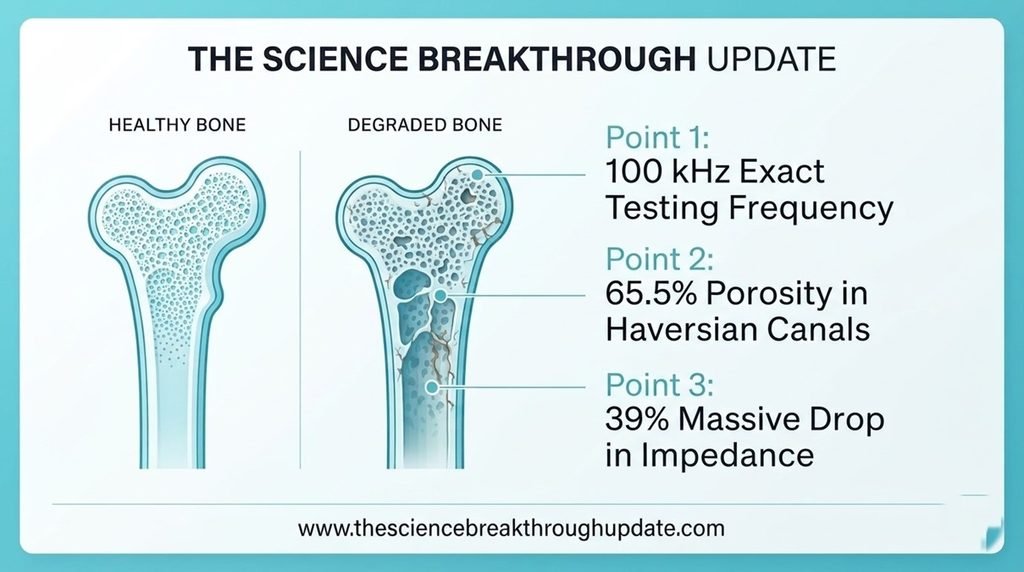
🧬 Researchers from Argentina successfully created a simplified multiscale computational model that accurately simulates how low-frequency electrical currents flow through living skeletal tissue. Their new approach captures the precise interactions between bone microstructure and effective conductivity at a specific frequency of 100 kHz.
📉 By combining advanced finite element simulations with mathematical homogenization models, the research team analyzed how structural changes alter macroscopic impedance. This work provides a vital physical foundation for developing future non-invasive diagnostic tools capable of detecting microstructural variations.
The Complex Challenge of Tracking Structural Heterogeneity
- 🔍 Current safety standards and electromagnetic exposure guidelines fail to account for the complex tissue heterogeneity and orthotropic anisotropy found across living skeletal structures.
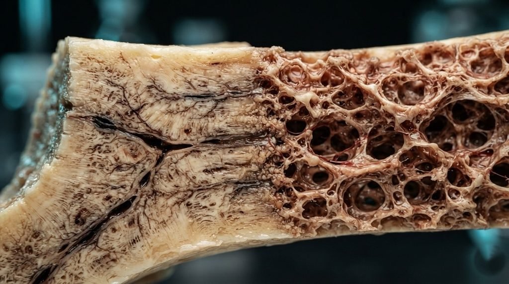
- 🔬 High-resolution imaging techniques like micro-computed tomography cannot fully resolve the dense, intricate networks of intracortical Haversian and Volkmann canals.
- 🎛️ Prior electrical homogenization frameworks rarely account for directional variations, making it difficult to assess how porosity levels alter overall current pathways.
- 📊 Medical researchers require a simplified, accurate method to connect microscopic tissue changes directly to the bulk electrical properties measured macroscopically.
Cracking the Code with Homogenization Rules
- 🧮 Corresponding author Ramiro M. Irastorza and the research team developed a multiscale approach combining finite element method simulations with Bruggeman-based models.
- 📈 The computational framework accurately captured the experimental conductivity range of 25 to 200 mS/m inside trabecular bone samples.
- 🦴 Structural analysis revealed that exactly 65.5% of the total porosity in the generated cortical geometries belongs to axial Haversian canals.
- 💻 The team integrated these microstructural fluid pathways into an anisotropic Bruggeman formulation to simulate directional current flow without heavy computational meshes.
Quantifying the Electrical Impact of Bone Loss
- 📉 Testing the properties on a macroscale bovine tibia model showed an approximate 38% to 39% reduction in total impedance for non-healthy bone.
- 💧 This significant drop in electrical resistance is driven by reduced trabecular bone volume fraction and a thinner outer cortical layer.
- ⚡ The simulations demonstrated that macroscale structural degradation reduces directional sensitivity, homogenizing the current paths across different electrode configurations.
- 🏥 These definitive results confirm that microstructural variations are clearly reflected in the macroscopic impedance of the simulated tibial model.
My Take: Replacing heavy scans with simple electrical math is a massive win. It lays the groundwork for cheap, everyday screening tools, finally moving bone diagnostics from expensive labs right into local clinics.
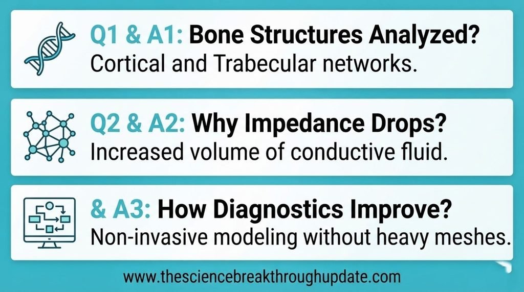
Reader Doubts: Core Questions Answered
- What specific bone structures were analyzed in this simulation?
- 🦴 The research analyzed two primary skeletal structural types known as cortical compact bone and trabecular cancellous bone. The cortical model explicitly incorporated a three-dimensional network of axial Haversian canals and transverse Volkmann canals. The trabecular model utilized real geometries derived from micro-computed tomography scans of bovine bone samples.
- Why does non-healthy bone exhibit a lower electrical impedance?
- 🔋 Non-healthy bone conditions involve a lower bone volume fraction and reduced cortical thickness, which increases the space available for conductive fluid. The higher presence of this physiological solution allows low-frequency electrical currents to pass through the tissue much more easily. Consequently, the overall macroscopic resistance drops significantly compared to healthy reference models.
- How do these electrical models help improve future medical diagnostics?
- 🛠️ These simplified mixing rules allow scientists to simulate complex skeletal structures without requiring massive, computationally heavy imaging datasets. The framework proves that surface electrical measurements are highly sensitive to overall structural degradation within the human body. This computational foundation assists engineers in designing more precise, non-invasive systems for monitoring internal skeletal changes.
Source Document Link: arXiv.org
Share this useful knowledge with your friends now!”
Disclaimer: The information on this website is for educational and informational purposes only and is not intended as medical advice. Always consult a qualified healthcare professional before making any medical decisions or changes to your treatment plan.